
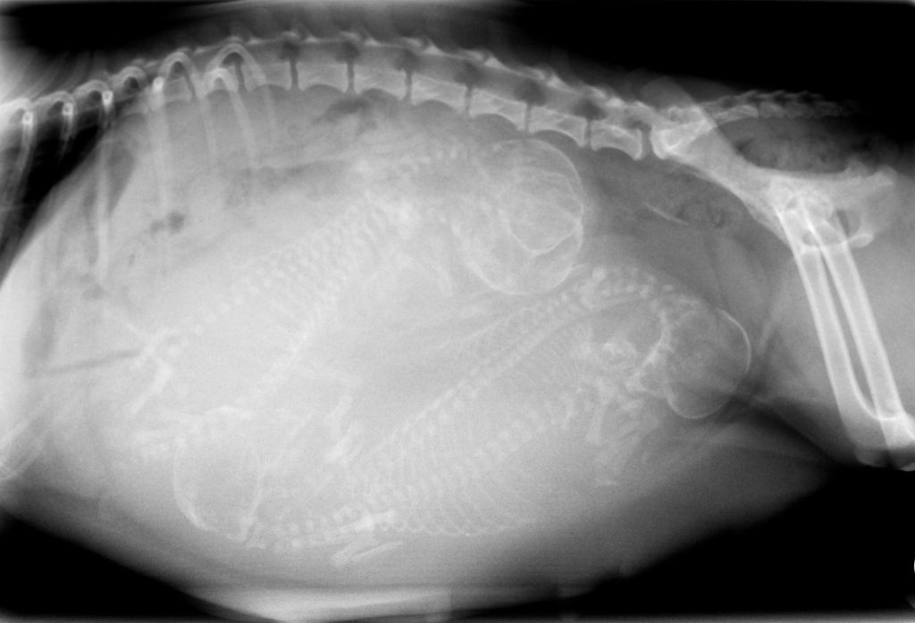
When a pet is sick or injured, an accurate diagnosis is an important first step toward determining the appropriate treatment. Springfield Animal Hospital uses diagnostic imaging to help veterinarians examine areas of the body that cannot be fully evaluated through a physical examination alone.
Request an AppointmentConditions Evaluated With Diagnostic Imaging
Diagnostic imaging can provide valuable information about your pet’s bones, joints, organs, and other internal structures. Depending on your pet’s symptoms and medical needs, imaging may help veterinarians evaluate injuries, abnormalities, and a variety of health conditions.
These diagnostic tools may be used to investigate concerns involving broken bones, joint problems, abnormal growths, spinal conditions, and internal organs such as the heart, lungs, liver, kidneys, bladder, stomach, and spleen.
Ultrasound Imaging
Ultrasound uses high-frequency sound waves to create images of structures inside your pet’s body. A handheld probe is gently moved over the area being examined, allowing the veterinarian to view internal organs and tissues on a monitor.
Ultrasound can be particularly helpful when evaluating organs such as the stomach, liver, spleen, pancreas, kidneys, and bladder. It may also assist in identifying abnormal masses, fluid accumulation, or foreign objects that a pet may have swallowed. Ultrasound is noninvasive and does not use ionizing radiation.
Radiographs
Radiographs, commonly called X-rays, provide images of structures inside your pet’s body. They are frequently used to evaluate bones and joints and can help identify fractures, changes in joint structure, and other skeletal abnormalities.
X-rays can also provide useful information about the chest and abdomen, including the size, shape, and position of certain internal organs. Depending on your pet’s symptoms, radiographs may be recommended as part of a broader diagnostic evaluation.
MRI
Magnetic Resonance Imaging, or MRI, produces highly detailed images of soft tissues and internal structures. This type of imaging can be especially useful when evaluating the brain, spinal cord, nerves, muscles, and other soft tissues.
MRI may be recommended when investigating certain neurological conditions, abnormal growths, or spinal problems. Because pets generally need to remain completely still during advanced imaging, sedation or anesthesia may be required.
CT Scans
Computed tomography, commonly known as a CT scan, uses X-ray technology to produce detailed cross-sectional images of the body. These images allow veterinarians to examine specific areas from multiple angles and can provide more detail than conventional radiographs in certain situations.
CT imaging may be useful when evaluating complex injuries, bones, internal organs, abnormal masses, and other conditions that require a more detailed view of your pet’s anatomy.
Diagnosing Your Pet’s Illness or Injury
The type of diagnostic imaging recommended will depend on your pet’s symptoms, medical history, physical examination, and individual needs. After reviewing the imaging results and other diagnostic findings, the veterinarian can discuss the diagnosis and recommend an appropriate treatment plan.
If your pet is experiencing pain, unusual behavior, mobility problems, or other concerning symptoms, Springfield Animal Hospital can help determine the next steps. The veterinary staff can also answer questions about diagnostic testing, treatment recommendations, and how to keep your pet comfortable during recovery.
For more information about radiology and diagnostic imaging services in El Segundo, call Springfield Animal Hospital at (904) 240-0426 today.